Leukoplakia causes white plaques & patches on the mucous membranes of the mouth.
Leuko stands for “white” and plakia for “patch”. Leukoplakia, classified as a potentially malignant disorder, is a white patch that cannot be rubbed of and cannot be clinically diagnosed as any other disease.
It has had numerous definitions over the years since it was first defined.
In 2005, WHO defined it as “a white plaque of questionable risk having excluded (other) known diseases or disorders that carry no increased risk for cancer”. The definition of leukoplakia is unusual in that the diagnosis is one of exclusion of other white lesions rather than depending on a definable appearance of the lesion.
Diagnosis of Oral Leukoplakia
To make it easier to understand, let’s say you have a patient with a suspicious white patch in the buccal mucosa.
Since several other white lesions look like that, make sure it is not any other white lesion.
Only after this, could you make a diagnosis of leukoplakia. Hence, it is a diagnosis of EXCLUSION!
Leukoplakia or any other white lesion for that matter appears white because of the thickened surface keratin layer and the hyperplastic epithelium.
This thickened abnormal keratin layer evenly reflects the visible light spectrum as opposed to being permeable to visible light
and the red spectrum being reflected by the connective tissue.
This clinically masks the vascularity (redness) of the underlying connective tissue making it appear white! Most cases of leukoplakia are related to the use of smoked or smokeless forms of tobacco and may or may not regress on discontinuation of the habits.
Other factors like alcohol, trauma, and candida Albicans have also been reported to cause leukoplakia although these associations are now being disputed.
Leukoplakia Causes
The worldwide prevalence of leukoplakia is believed to be around 2% and it increases with age.
It was known to have a higher male predilection but is now seeing a shift with a nearly equal number of women being affected.
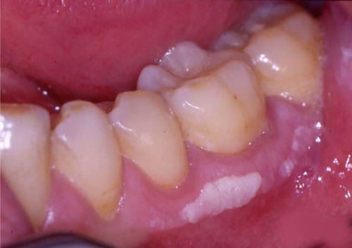
Buccal mucosa, mandibular vestibule, gingiva, and lateral borders of the tongue are most commonly affected.
Other sites though rare include, the lip, the floor of the mouth, retromolar trigone, palate, mandibular and maxillary alveolar ridges.
Different Types of Leukoplakia
Leukoplakia is classified as being homogenous and non-homogenous.
Homogenous refers to the uniformity of the color and the texture of the leukoplakic lesion while non-homogenous
leukoplakia may have a red-white appearance or may have a uniform white appearance with an irregular texture manifesting with nodules wart-like projections and corrugations.
Homogenous leukoplakia in the early stages may be thin, flat, or slightly elevated, grey-white in color with fissures. While the leukoplakia that has progressed could be a thicker white plaque with deep fissures.
These lesions are well-circumscribed with sharply demarcated borders.
If these homogenous lesions develop surface irregularities,
they are called granular or nodular leukoplakias.
They are called verrucous leukoplakias when they develop deep corrugations and wart-like projections.
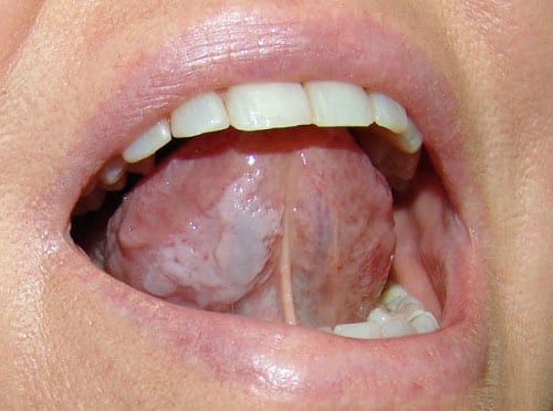
Leukoplakia may reveal red patches manifesting as a mixed red-white lesion, called erythro-leukoplakia or speckled leukoplakia.
A notable form of leukoplakia, called proliferative verrucous leukoplakia, appears in multiple sites showing slow, persistent growth eventually becoming verrucous and exophytic with a high rate of recurrence even after treatment.
How Herpes Is Diagnosed?
Herpes can usually be diagnosed based on how the skin or mucous membrane lesions look and can be confirmed with tests looking for viral DNA, like a polymerase chain reaction, an antibody response to the virus, or by growing the virus with viral culture.
Although infections typically resolve without treatment within a couple of weeks, there are antiviral drugs like acyclovir, famciclovir, and valacyclovir that can be used topically or systemically to reduce pain and speed up healing.
For recurring episodes, these treatments usually work best if taken when the prodrome starts; in other words, before the blisters develop, and high-dose intravenous antivirals may be given in more severe, or life-threatening situations.
Prognosis of Leukoplakia
The prognosis of leukoplakia depends on various factors and is generally considered bad If it manifests in males if the lesion is present for a long duration if the affected is a non-smoker if leukoplakia is located on the tongue or floor of the mouth if the size is greater than 2 cm and the lesion is non-homogenous In the absence of dysplasia, excision is not mandatory although the patient should be under continuous surveillance and evaluation every 6 months.
Re-biopsy of suspicious areas should be done whenever necessary. The use of antioxidant nutrients like Vitamin A, C, and E, beta carotene, and a diet high in antioxidants – fruits and vegetables are recommended.
Leukoplakia with mild dysplasia is managed according to its size and response to conservative measures like habit cessation.
For moderate to severe dysplasias, excision should be done if feasible, although there are several studies that report that surgical excision does not reduce the risk of recurrence of the patient developing carcinoma.
Grafting procedures may be necessary after large excisions.
Two groups of orthodontic treatments
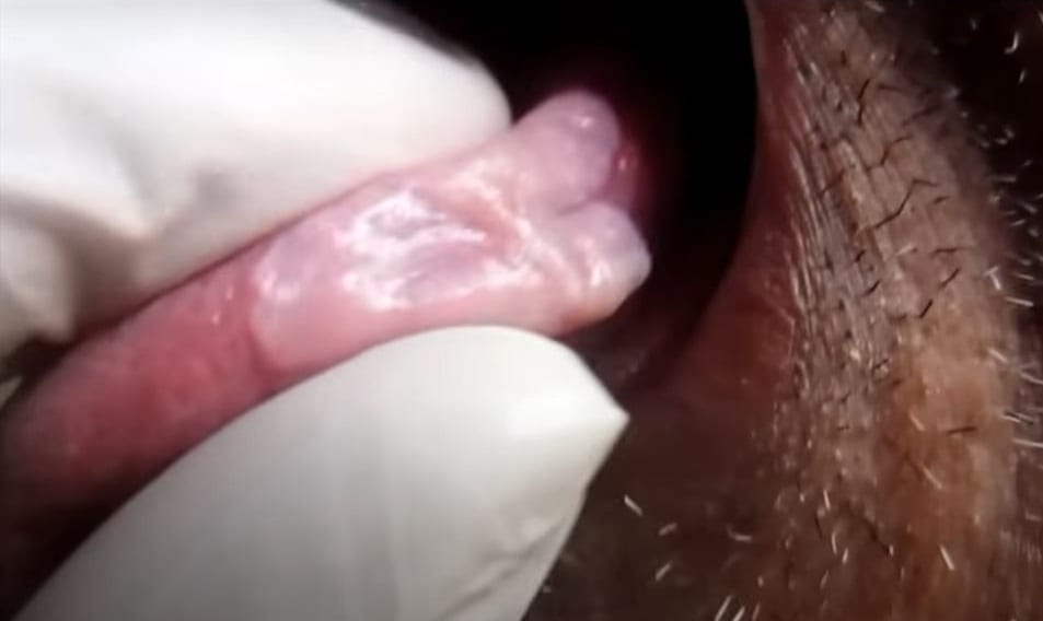
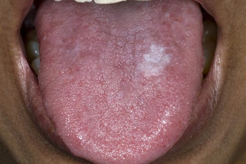
You can see the whitish lesion on the lateral side of the tongue with a fissure in the center on the palpation. It was giving a very dry and leathery feel that appears like homogeneous leukoplakia.
The Cretan layer becomes thick that hyper pyrolysis happens. It appears white when get hydrated with the saliva. He has a positive history of tobacco chewing for about 40 years. This could be confirmed by a biopsy of what is.